

Introduction
A level 3 probabilistic safety assessment (PSA) aims to estimate the environmental and health effects of radiation from nuclear accidents on the public. To this end, various codes have been developed for level 3 PSA: in the U.S., this includes the MELCOR accident consequence code system (MACCS) developed by the Nuclear Regulatory Commission (NRC); in Japan, the off-site consequence analysis of atmospheric releases of radionuclides (OSCAAR); and in the European Commission (EC), the code system from MARIA (COSYMA) [1]. However, there is no level 3 PSA code unique to Korea, and the U.S. NRC code MACCS has been adopted for level 3 PSA.
After the Fukushima nuclear accident in 2011, there has been increasing demand for off-site consequence analyses in terms of the environmental and health impacts of radiation exposure. In Korea, the recent enactment of a new nuclear safety act has obligated relevant organizations to conduct a level 3 PSA, increasing the need to develop a level 3 PSA code specific to Korea.
A level 3 PSA sequentially evaluates the atmospheric dispersion of radioactive material, exposure dose, and public health effects. As well as being an integral component of off-site consequence analyses, assessing the impact of radiation on public health involves quantification of these health effects. Radiation-induced public health effects can be classified as short-term or long-term effects. Short-term effects occur when the body exposed to a large amount of radiation. These effects reveal themselves within a year of exposure and are regarded as deterministic effects associated with exposure more than a threshold dose of radiation. Long-term effects surface several decades after the radiation exposure. They are probabilistic effects that occur without a threshold dose. Examples of long-term effects of radiation are cancer induced by somatic mutations and genetic (i.e., heritable) diseases induced by mutations in reproductive cells [2]. As seen in post-nuclear accidents, chronic exposure is thought to cause long-term health effects, generally cancer; thus, parameters such as the incidence and mortality of cancer are important scales when assessing radiation-induced public health impacts. Accordingly, multiple organizations have developed models estimating radiation-induced cancer incidence and/or mortality, and recently these cancer risk models have been refined using the most recent updates of atomic bomb survivor data from Japan.
In 1972, the U.S. National Academy of Sciences published its initial report on the biological effects of ionizing radiation (BEIR) and developed a risk model for radiation-induced cancer [3]. In its seventh publication, the BEIR VII report in 2006, a new risk model was presented that reflects the latest data from the Japanese atomic bomb database [4, 5]. Similarly, in their 1993 NUREG/CR-4214 report, the U.S. NRC published an updated cancer risk model for exposure to radiation from nuclear accident, which is a revision of the initial model they published in 1985. Later, the U.S. NRC developed an MACCS code as a tool for level 3 PSA, which is built upon the cancer risk model published in NUREG/CR-4214 in 1990 [6ŌĆō11]. The United Nations Scientific Committee on the Effects of Atomic Radiation (UNSCEAR) and the International Commission on Radiological Protection (ICRP) have each developed an updated cancer risk model, which were presented in the UNSCEAR 2006 report and the ICRP 103, respectively [12, 13]. Other models have also been developed and used, for instance by the U.S. Environmental Protection Agency (EPA), which created an improved version of the BEIR VII (2006) model, and by the World Health Organization, which adapted the latest models of UNSCEAR and ICRP to conduct off-site consequence analyses of the Fukushima nuclear accident [14, 15]. These reports indicate that multiple organizations are directing their efforts and resources to develop and enhance models to determine the health effects of radiation and to estimate the associated risks, such as radiation-induced cancer risk.
In advance of developing a model of health risks specific to the Korean population, and thereby a level 3 PSA code specific to Korea, we evaluated the latest cancer risk models of different organizations. We analyzed the methodological approaches employed by each organization to develop these cancer risk models and compared the epidemiological data, dose-response functions, and the selection of cancers across organizations. Recommendations based on our findings are presented with respect to the development of cancer risk models specific to the Korean population.
Materials and Methods
1. Mathematical modeling
The risk of cancer due to radiation can be defined using both multiplicative and additive models. The multiplicative model assumes that radiation-induced excess cancer risk shows a relative increase in risk, or excess relative risk (ERR), in proportion to the baseline rate. The additive model assumes that radiation-induced excess cancer risk shows an absolute increase in risk, or excess absolute risk (EAR) associated with exposure independently of the baseline rate. The radiation-induced excess cancer risk is calculated using the following equations:
(3)
Here, M(D,e,a,g) denotes radiation-induced excess cancer risk, with the following functions: dose of exposure (D), age at exposure to radiation (e), attained age of an individual (a), and gender (g). Additionally, ╬╗u (a,g) denotes cancer risk without exposure to radiation.
Simply, both ERR and EAR are calculated as the dose-response function, Žü(D), multiplied by the effect modification, ŽĄ(X). The basic formula of Žü(D) is a linear, linear-quadratic, or quadratic form and ŽĄ(X) has an exponential form with respect to variables such as age at exposure, attained age, and gender. The coefficients of the equation are determined by conducting a regression analysis of the epidemiological data (e.g., of atomic bomb survivor data and medical records/databases) of the exposed population, which are then added to the basic formula of Žü(D) and ŽĄ(X) [16, 17].
dose-response function,
Žü ( D ) = { ╬▒ D ╬▒ D + ╬▓ D 2 ╬▓ D 2
effect modification, ŽĄ(X)=e╬ĖŌĆ▓X, ╬ĖŌĆ▓=variable factor
The term Žü(D) describes dose-dependent cancer risk, whereas ŽĄ(X) adjusts for cancer risk attributed to non-radiological factors such as age at exposure, attained age, and gender under the same dose exposure. Estimates of low-dose radiation-induced cancer risk are extrapolated from risk models derived from high-dose exposure data, such as the Japanese atomic bomb survivor data. Because the cancer risk in the range of low dose may be overestimated in the linear Žü(D) model, the dose and dose rate effectiveness factor (DDREF) has been used to adjust for this.
2. NUREG model (1993)
The NUREG model assumes that Žü(D) is linear. The DDREF is used for exposure to radiation doses below 0.2 Gy or dose rates below 0.1 Gy┬ĘhŌłÆ1. The term ŽĄ(X) is not defined in this model, and risk is adjusted using age group-specific risk coefficients. The excess cancer risk model is shown in Table 1. Further, the cancer risk estimates of the NUREG model use risk coefficients defined in terms of cancer risk per unit of dose calculated in previous studies, such as the BEIR III study.
cŽä=risk coefficient by age group (GyŌłÆ1)
The lifetime cancer risk of the population is calculated by multiplying the excess cancer risk over a specific period of time after the initial radiation by population-specific characteristics, such as the distribution of population and survival rates by age group.
Here, R(Žä,D) denotes the mean cancer risk from exposure to radiation over a certain period of time after the initial radiation (Žä). The population-specific factors fk and Sk (Žä) denote the population distribution by age group (k) and the survival rate of the age group (k) during this time (Žä), respectively [9]. Because the MACCS code, which is the adopted code in Korea, calculates lifetime cancer risk using an R(D) equation based on data from the US National Census in 1980, estimating the cancer risk in Korean populations with the MACCS code may lead to errors [10].
3. BEIR VII model (2006)
The BEIR VII model is developed based on epidemiological data from radiation-exposed populations, and aims to estimate cancer risk induced by exposure to low-linear energy transfer radiation. Risk models for cancer incidence and mortality have been developed for leukemia and for solid cancers. Models for solid cancers were further developed according to site (stomach, colon, liver, lung, breast, prostate, ovary, bladder, thyroid, and others). The risk model for leukemia was derived from the Japanese atomic bomb mortality data from the period 1950ŌĆō2000 [18], and that for the solid cancers from the Japanese atomic bomb cancer incidence data from the period 1958ŌĆō1998 [17]. Cancer risks are predicted as weighted means of the ERR and EAR estimates and adjusted for age at exposure, attained age, gender, and time after initial exposure (t). The leukemia model has a linear-exponential Žü(D) term and a latency period of 2 years. The solid cancer model has a linear Žü(D) term with an estimated DDREF of 1.5 and a latency period of 5 years. The ERR and EAR models are shown below, and the coefficients of the BEIR VII model are presented in Table 2.
Although the equations for ERR and EAR are identical for risk models of cancer incidence and mortality, the risk of cancer mortality is calculated by multiplying the risk of cancer incidence by the ratio of the baseline rate of cancer morality, ╬╗m (a) to the baseline rate of cancer incidence of the unexposed population, ╬╗i (a).
Excess cancer incidence risk:
Excess cancer mortality risk:
The lifetime risk of cancer of the exposed population is calculated using the lifetime attributable risk (LAR), which describes the accumulated risk from the initial radiation. When calculating the LAR of solid cancers, excluding thyroid and breast cancers, and of leukemia, the weighted geometric mean is utilized (L=latency, S(a)=survival rate by age group of the study group) [5].
4. UNSCEAR model (2006)
Risk models for the incidence and mortality associated with radiation exposure were developed for leukemia and solid cancers by UNSCEAR. These cancer risk models were derived through regression analyses of the Japanese atomic bomb survivor cancer mortality data from the period 1950ŌĆō2000 and of Japanese atomic bomb survivor cancer incidence data from the period 1958ŌĆō1998. The risk models for mortality from leukemia and solid cancers have a linear-quadratic Žü(D) term, and the sites of solid cancers are not specified. The risk model for the incidence of solid cancers is linear and was developed for various cancer sites (Table 3). Only the Žü(D) of risk models for incidence of bone cancer and non-melanoma skin cancer have a linear-quadratic function and a quadratic-exponential function, respectively. For leukemia, the risk models for cancer incidence and mortality are the same. The DDREF was presented as 2 and the ERR and EAR estimates were adjusted via ŽĄ(X) with respect to age at exposure, attained age, and gender.
Risk model for the incidence of and mortality from leukemia:
╬▒ = 864.55 SvŌłÆ1,
╬▓ ╬▒ = 1.18 ŌĆē Sv - 1
╬▒ = 7.52 ├Ś 10ŌłÆ4 SvŌłÆ1,
╬▓ ╬▒ = 1.04 ŌĆē Sv - 1
Risk model of mortality from solid cancers:
╬▒ = 408.29 SvŌłÆ1,
╬▓ ╬▒ = 0.29 ŌĆē Sv - 1
╬▒ = 7.75 ├Ś 10ŌłÆ9 SvŌłÆ1,
╬▓ ╬▒ = 0.40 ŌĆē Sv - 1
Risk model for incidence of solid cancers:
(19)
The lifetime risk of cancer of the population is derived from parameters such as excess cancer death and risk of exposure-induced death [12].
5. ICRP 103 model (2007)
The ICRP 103 presents models for cancer incidence. With improved outcomes of cancer treatment, radiation-induced health effects are emphasized in connection with the incidence of cancer, instead of cancer-related mortality. The model for leukemia incidence is based on the Japanese atomic bomb survivor data from the period 1950ŌĆō1987 [19], and the model for solid cancer incidence is based on the Japanese atomic bomb survivor data from the period 1958ŌĆō1998. It is assumed that the Žü(D) term is linear-quadratic for leukemia and linear for solid cancers (with a DDREF of 2). The lifetime risk for cancer is calculated using the LAR and the weighted arithmetic mean, using the LARs of ERR and EAR. In the ICRP 103 model, solid cancers are divided into the following sites: thyroid, breast, esophagus, stomach, colon, liver, lung, ovary, bladder, and others. The two models estimating cancer incidence are shown below, and their coefficients are presented in Table 4 and 5 [13].
6. U.S. EPA model (2011)
The U.S. EPA developed risk models for cancer incidence and mortality, using the same models as the BEIR VII model. Additionally, they developed new risk models for thyroid cancer and mathematical models for kidney, bone, and skin cancers. It is assumed that the cancer risk model for thyroid cancer has a linear Žü(D) term, and this model is defined using ŽĄ(X) related to age at exposure and time after exposure. The cancer risk model for kidney cancer is defined as the EAR per unit of dose by multiplying the solid cancer model of the BEIR VII model by the incidence rate of kidney cancer, ╬╗i,kidney, divided by the incidence rate of other solid cancers, ╬╗i,residual. The cancer risk model for bone cancer is based on statistics on alpha radiation-induced bone cancer and for skin cancer, on tinea capitis cohort data.
Thyroid cancer:
╬▒ = 10.7 GyŌłÆ1,
A { 1.0 , e < 5 0.6 , 5 Ōēż e Ōēż 9 0.2 , 10 Ōēż e Ōēż 14 , 0.2 e - 0.083 ŌĆē ( e - 15 ) , 15 Ōēż e
Kidney cancer:
Bone cancer:
g(e) = eŌłÆ0.0532(eŌłÆ30),
╬▒ = 178 ├Ś 10ŌłÆ3 GyŌłÆ1, t0 = 12.72, Žā = 0.61
The lifetime risk of cancer in the radiation-exposed population is calculated using LAR, as in the BEIR VII model. Although the BEIR VII model sets the upper range of age at 100 years, the U.S. EPA model sets it to 110 years and computes the risk as a weighted arithmetic mean of the ERR and EAR estimates [14].
Results and Discussion
In advance of the development of a Korean health risk assessment model, we examined the preexisting cancer risk models used by nuclear- and radiation-related organizations worldwide. With improved cancer treatment rates, radiation-derived cancer risk generally refers to the risk of cancer incidence rather than the risk of cancer mortality. Thus, each organization analyzed in this study has developed risk models for cancer incidence as well as cancer mortality. Current mathematical models for cancer risk distinguish the risk of leukemia from that of solid cancers. Furthermore, risk models for solid cancers specify the site of cancer, as shown in Table 6. Of the risk models, the U.S. EPA model describes the most number of models for cancer (15 cancers, excluding only esophageal cancer and brain and central nervous system [CNS]-related cancers). Table 7 compares the risk models by organization. These models can be categorized into 2 groups according to the methodology of how they were developed: (1) those based on epidemiological data to develop ERR and EAR (the BEIR VII, UNSCEAR, ICRP, and U.S. EPA models), and (2) the NUREG model, which uses risk coefficients derived from preexisting studies and determines radiation-induced cancer risk by multiplying these risk coefficients by the exposure dose. Additionally, the BEIR VII UNSCEAR, ICRP, and U.S. EPA models use a modification function to adjust for the effects of age at exposure, attained age, and gender on cancer risk under the same exposure conditions, but the NUREG model does not. Instead, modification calculations are possible using a set of different risk coefficients that can be applied to different subgroups of gender and age.
The epidemiological data that have been applied to the BEIR VII, the UNSCEAR, the ICRP, and the U.S. EPA models are mostly derived from Japanese atomic bomb survivor data. From this dataset, the cancer mortality data until 2000 and the cancer incidence data until 1998 were used. The models also include epidemiological finding incorporating medical exposure data, although to a lesser extent. Thus, the resulting model may differ depending on the combination of other radiation exposure data used, even if the major epidemiological data in all models are the Japanese atomic bomb survivor data.
Ideally, risk models should be developed using arbitrarily sampled epidemiological data from radiation exposure data that are not derived from specific nations or populations. However, currently available large-scale radiation exposure data are restricted to data from a specific population (i.e., the Japanese atomic bomb survivors); therefore, applying risk models derived from such country/population-specific data to other countries or populations can lead to errors. Thus, in numerous organizations, geometric and arithmetic means, which take into account the weighted factors of ERR and EAR, are used to offset the potential errors. Despite these precautions, limitations and uncertainties associated with the epidemiological data, which are currently heavily restricted to the Japanese population, still remain, and the Korean cancer risk model will share the same difficulties. Further, the Japanese atomic bomb survivor data generally reflect acute, high-dose exposure to high-energy radiation, meaning that estimating the risk associated with low-dose and low-dose-rate radiation and different types of radiation may be difficult.
The DDREF, initially adopted by the ICRP, complements the limitations of the high dose and dose-rate exposure data and allows the estimation of risk from low dose and dose-rate exposures. These DDREF values are derived from life span study data and from animal experiment findings. Although the use of DDREF is a simple and practical way of estimating low dose and dose-rate risk, the effectiveness of DDREF for extrapolating risk from the high dose and high dose-rate exposure effects is still under question. Representative cell biology findings that purport to support the DDREF are vague and obscure, and whether the findings of animal experiments can be translated to humans remains debatable. Recently, the German Radiation Protection Committee argued that the use of the DDREF can no longer be justified [20]. Thus, prior to developing a Korean risk model, an evaluation of the limitations of the Japanese atomic bomb survivor data and of the effectiveness of the DDREF should be carried out.
Through the comparison of the current cancer risk models, we analyzed the factors that must be taken account when developing a Korean cancer risk model. First, the risk models that have been developed calculate the risks for the following 17 types of cancers: leukemia, thyroid cancer, breast cancer, stomach cancer, colon cancer, liver cancer, lung cancer, prostate cancer, uterine cancer, ovarian cancer, bladder cancer, renal cancer, bone cancer, skin cancer, esophagus cancer, brain and CNS-related cancers, and other solid cancers. The Korean cancer risk model should include the most prevalent cancers among the Korean population. According to the 2013 national cancer incidence statistics, cancers with a 5-year cancer incidence greater than 3% included solid cancers in regions including the thyroid, stomach, large intestine, lung, breast, liver, and prostate. Thus, the Korean cancer risk model should include these cancers, as well as leukemia.
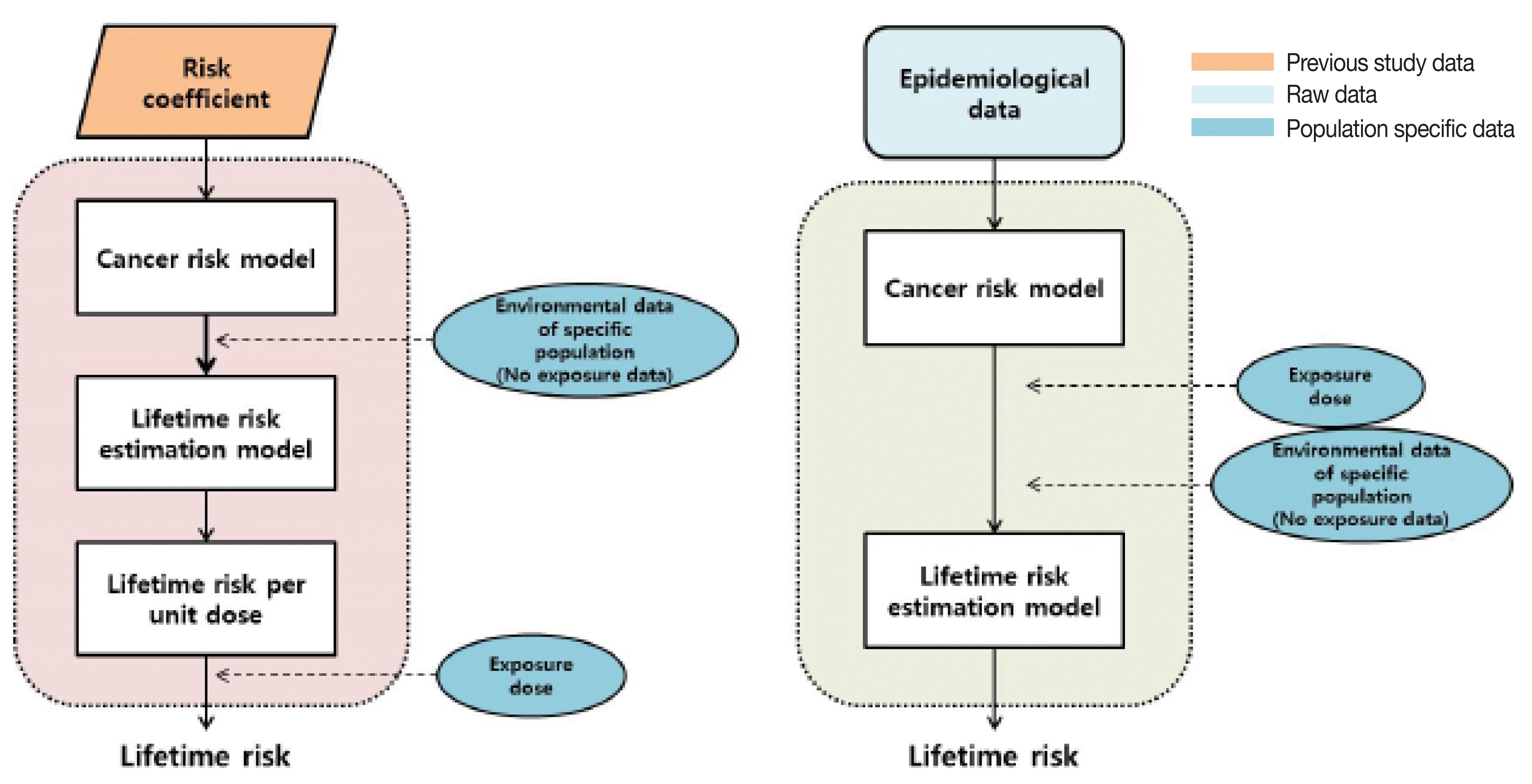
Second, we make methodological suggestions for the development of a Korean risk model. As shown in Figure 1, pre-determined risk coefficients were used to develop the NUREG model, along with unexposed population data, to calculate the lifetime risk per unit dose. The BEIR VII, UNSCEAR, ICRP, and U.S. EPA models were derived from epidemiological data. These models calculate lifetime risk by age at exposure, attained age, and gender. Both methodologies provide advantages when developing a Korean risk model: the risk coefficient-derived methodology is a relatively simplified process of creating a mathematical model, while the epidemiological data-based methodology allows the latest data to be incorporated. Further, because both methods use unexposed population data, national census data can be used, allowing the contextualization of the model to the Korean characteristics. Further, because the development of the Korean risk model will employ the latest datasets of the Japanese atomic bomb survivor data (up to 2009), in contrast to the current risk models that have used data up to 2000, we will be able to create the most up-to-date risk model. Thus, the methodology of the Korean risk model development should prioritize collecting and using the latest Japanese atomic bomb survivor data.
Finally, each organization applied a different DDREF value, meaning that a DDREF value for the Korean risk model is also needed. We suggest that this DDREF value should be determined on the basis of recent studies regarding low dose and low-dose-rate radiation exposure-dependent cancer risk. Because studies evaluating the effectiveness of the DDREF have not been extensively conducted in Korea, future studies are required concerning this widely employed factor.
Conclusion
In advance of the development of a health risk assessment model specific to the Korean population, and thereby a level 3 PSA code specific to Korea, we evaluated the latest cancer risk models used by organizations worldwide. The following cancer risk models were evaluated: the NUREG model (1993), the BEIR VII model (2006), the UNSCEAR model (2006), the ICRP 103 model (2007), and the U.S. EPA model (2011). In this study, the methodological approaches and the epidemiological data of these models were comparatively analyzed, and the different cancer sites of each model were summarized.
Through a comparative analysis of existing cancer risk models, we determined important factors that must be considered in order to develop a Korean cancer risk model. We suggest that the Korean cancer risk model should be developed on the basis of the latest epidemiological data and should include the following key cancer sites: the thyroid, stomach, large intestines, lung, breast, liver, prostate, and other solid cancers, as well as leukemia. These cancer sites are suggested on the basis of the cancer incidence statistics data in Korea from the previous 5 years. In order to develop an up-to-date model, we also suggest that the Japanese atomic bomb survival data for the period up to 2009 should be used. In conclusion, the findings of this study are expected to serve as the basis for future studies on the development of a Korean cancer risk model.