

Introduction
Generally, the response of neutron measuring instruments used for radiation protection strongly depend on incident neutron energy. Therefore, for the reliable measurement using such instruments, those should be calibrated at the calibration field whose neutron spectrum is similar to an actual workplace. RI neutron sources, such as 252Cf or 241Am-Be sources, are often used for calibration of neutron measuring instruments, however, neutron spectra of such RI sources are significantly different from those at actual workplaces in most cases. Thus, moderated neutron calibration fields that have continuous neutron spectra simulating actual workplaces are required.
At the Facility of Radiation Standard (FRS) in the Japan Atomic Energy Agency (JAEA), concrete-moderated neutron fields [1] using an 241Am-Be source were once used for calibration of neutron survey meters. However, it was greatly damaged by the Great East Japan Earthquake in 2011 and became unavailable. Therefore, we developed new moderated neutron calibration fields using a graphite moderator and 241Am-Be sources based on ISO12789 [2, 3] which provides a guideline for producing and characterizing simulated workplace neutron fields. In this paper, we describe the characteristic of our new calibration fields.
Graphite-moderated Neutron Calibration Fields
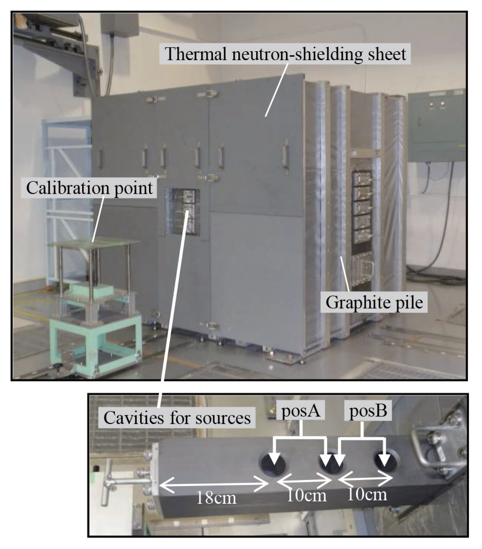
The overview of new moderated neutron calibration fields is shown in Figure 1. It was constructed at the corner of the neutron irradiation room of 12.5 m ├Ś 12.5 m ├Ś 11.7 m in the FRS. The fields use a graphite pile (150 cm(W) ├Ś164 cm(D) ├Ś150 cm(H)) as a moderator, which is also used for the thermal neutron calibration field [4] in the FRS. While a 252Cf source is installed into the cavity at the center of the graphite pile for the thermal neutron field, two 241Am-Be neutron sources (37 GBq├Ś2) are installed into cavities located at the shallower position inside the graphite pile. By using two 241Am-Be sources, enough dose equivalent rates for calibration are obtained. Two calibration fields with different neutron spectra are available by changing positions of neutron sources; posA: sources at 18 cm and 28 cm in depth from surface, posB: sources at 28 cm and 38 cm in depth from surface, as shown in the lower picture in Figure 1. Furthermore, the thermal neutron-shielding sheet including gadolinium is put on the surface of the graphite pile, to decrease an amount of thermal neutrons at the calibration point. At the center of the sheet, there is an aperture of 30 cm ├Ś 30 cm for putting in or taking off sources. The calibration point is fixed at 75 cm from the surface of the graphite pile, and 81.5 cm above the floor as the same height as sources.
Neutron Spectra and Reference Values
First, we calculated neutron energy spectra of the fields using the Monte-Carlo simulation code MCNP-5 [5] with a library of nuclear data JENDL3.2 [6]. The volume tally (F4), a sphere with radius of 5 cm, was used for accumulation of neutron events. Not only the graphite moderator and the thermal neutron-shielding sheet, but also capsules of sources, floor, ceiling and walls of the irradiation room, which are possibly affect a neutron spectrum at the calibration points, were considered into the geometrical model for calculations. The composition of impurities in the graphite was considered with referring data sheet summarized in the literature [4]. The energy spectrum of neutrons emitted from 241Am-Be source was taken from ISO8529 part1 [7].
Then, we evaluated neutron energy spectra at the calibration point, using the Bonner Multi-sphere Spectrometer (BMS). The BMS consists of a spherical BF3ŌĆōfilled proportional counter (5.1 cm in diameter, 0.26 atm) and polyethylene moderators with 8 different thicknesses (1, 2, 3, 4, 6, 8, 10, and 14 cm in thick). The BMS used here was well-calibrated at 252Cf and mono-energetic neutron calibration fields in the FRS. Thermal neutron fluence was measured separately by the cadmium difference method using the bare BF3-filled counter with/without a cadmium cover. The spectra were unfolded using SANDII [8] code, in conjunction with initial guess spectra calculated with MCNP code.
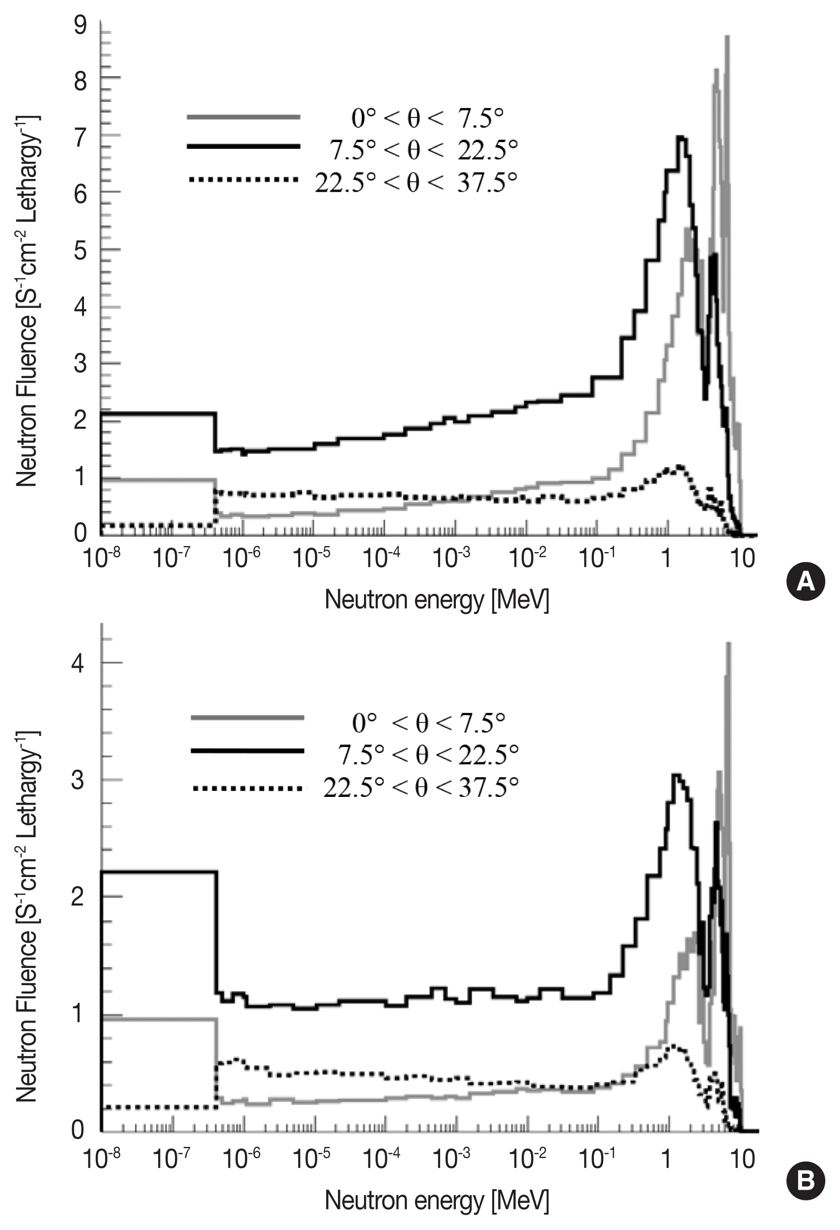
Neutron energy spectra for posA and posB by calculations and measurements were shown in Figure 2. Continuous neutron spectra in the wide energy range from thermal neutron energy to several MeV were given. The spectrum for posB was flatter than that for posA, because of longer path length in the graphite. Scattered neutrons make a large peak around 2 MeV, which is similar to a neutron spectrum of bare 252Cf source. And highly scattered neutrons make a continuous tail in the epi-thermal and thermal energy region. The ratios of epi-thermal neutron (0.414 eV-150 keV) fluence for total fluence are 47% and 48% for posA and posB, respectively. The spectra by measurement and calculation were approximately consistent.
Reference ambient dose equivalent rate H*(10) and personal dose equivalent rate Hp(10) were determined from neutron energy spectra measured using the BMS, as below,
where ╬”(E) is neutron fluence, h*(10;E) and hp(10;E,╬Ė) are conversion coefficients from neutron fluence to dose equivalent, taken from ICRP 74 [9]. The fluence for each incident angles ╬Ė was not evaluated experimentally, therefore parallel rays (that is, ╬Ė=0) was assumed to calculate Hp(10). The relevance to assume parallel rays is discussed in the section 5. In the energy region of thermal neutron (<0.414 eV), the energy distribution is assumed to be Maxwell-Boltzmann distribution with kT=0.025 eV. The Reference values of fluence rates, dose equivalent rates and averaged energies of the fields were summarized in Table 1. The given fluence and dose equivalent rates are enough for calibration of neutron measuring instruments. The H*(10) and Hp(10) values agreed with those determined from calculated neutron spectra within 4%.
Uniformity of Irradiation
We evaluated the spatial uniformity of dose equivalent rates in the irradiation field, by the Monte Carlo calculations. Dose rates were calculated using a point tally (F5) of MCNP, by changing position of test point along horizontal direction (X) and vertical direction (Z). The dose rates at each position were normalized by the value at the calibration point (that is, X=Z=0). As shown in Figure 3, the dose rates were found to be uniform within 5% in the area of ┬▒15 cm from the calibration point, for both directions of X and Z. The uniformity was confirmed in the sufficient area to calibrate neutron survey meters or personal dosimeters fixed on the phantom. The dose rate at the calibration point is slightly lower than those at surrounded areas, because the drawer pull located between sources and the calibration point disturbs incidence of direct neutron from primary sources.
Distribution of Incident Angle
In the previous section, though Hp(10) value depends on the incident angle of neutrons, we assumed parallel neutron incidence to calculate it. We next evaluated an angular distribution of incident neutrons at the calibration point with the Monte Carlo calculations, in order to verify whether the assumption of parallel rays is appropriate or not. We used a surface current tally (FSn) of MCNP. The surface tally of 5cm in radius was set to the calibration point with facing the graphite pile. The incident angle ╬Ė was defined as an angle between direction of incident neutrons and the line connecting the calibration point and sources; that is, an incident angle of uncollided neutrons is ╬Ė=0┬░.
The energy spectra of neutrons coming from angular intervals of 0┬░<╬Ė<7.5┬░, 7.5┬░<╬Ė<22.5┬░, and 22.5┬░<╬Ė<37.5┬░ are shown in Figure 4. The spectral shapes were found to be significantly different by incident angle. In the interval of 0┬░<╬Ė<7.5┬░, uncollided and lowly scattered neutrons are dominant. On the other hand, in intervals of 7.5┬░<╬Ė<22.5┬░ and 22.5┬░<╬Ė<37.5┬░, an amount of multiply-scattered neutrons in the epi-thermal energy region increases. Note that thermal neutrons coming from the interval of 22.5┬░<╬Ė<37.5┬░ were mostly cut due to the thermal neutron-shielding sheet. The spectra of neutrons coming from ╬Ė>37.5┬░ were not shown in the figure, because the fraction of those neutrons was less than 4% of total fluence.
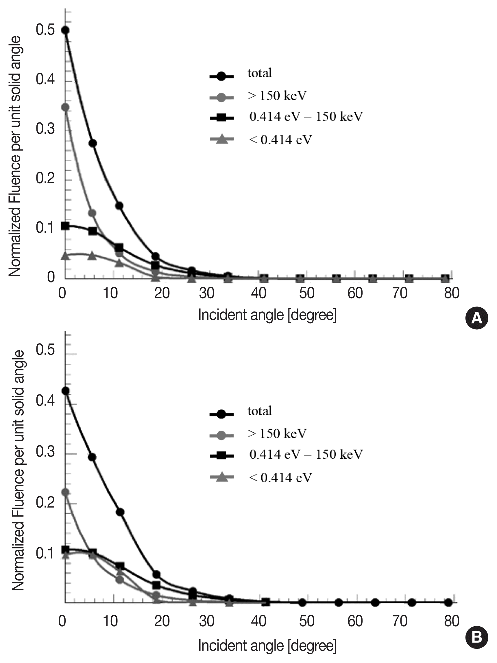
The angular distribution of neutron fluence per unit solid angle are shown in Figure 5, with dividing into three energy ranges; a high energy component (>150 keV), an epi-thermal component (0.414 eV-150 keV) and a thermal component (<0.414 eV). We found that almost all neutrons come with small incident angles less than 30┬░. Then, we calculated Hp(10) rate using formula written in section 3, with considering angular distribution of incident neutrons. The Hp(10) rates for posA and posB were given as 50.7 ╬╝Sv┬ĘhŌłÆ1 and 22.0 ╬╝Sv┬ĘhŌłÆ1, respectively; those values are only 1ŌĆō2% less than reference Hp(10) rates summarized in Table 1. Therefore, we confirmed that the assumption of parallel rays to calculate Hp(10) was appropriate.
Conclusions
The graphite-moderated neutron calibration fields using two 241Am-Be sources were constructed in the JAEA-FRS. Reference values of fluence rates, dose equivalent rates of H*(10), Hp(10) and averaged energies of two fields (posA and posB) were determined from neutron spectra measured by BMS. The reference Hp(10) values, which were determined with assuming parallel neutron rays, were only 1ŌĆō2% less than those calculated with considering angular distribution of incident neutrons. Currently, we are able to provide both H*(10) and Hp(10) values, for calibration or performance test of neutron measuring instruments.